
Intraoral Cameras
Intraoral Cameras
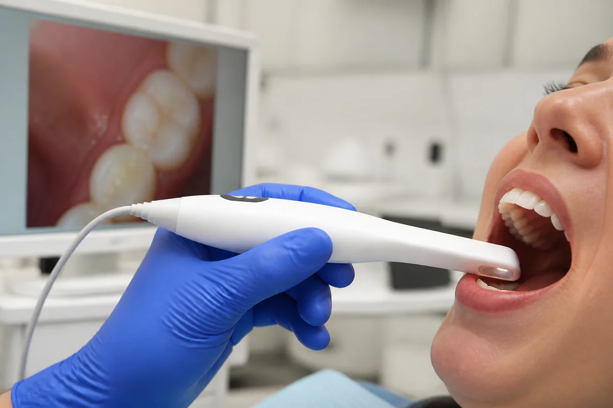
An intraoral camera is a small, pen-sized camera used during a dental exam to capture close-up photos of your teeth and gums. At Dental Office of Dr. Salm, Kanner, and Grandeau, these chairside cameras let patients see photos of their own teeth in real time, making it easier to understand what the dentist sees and why a particular treatment might be recommended.
What intraoral Cameras is
An intraoral camera is a compact wand fitted with a lens and a light source, connected to a computer monitor in the treatment room. As the dentist or hygienist moves it gently around the mouth, it captures magnified images of individual teeth, fillings, gum tissue, or areas of concern, displaying them instantly on a screen the patient can also view.
Before tools like this became common, patients typically had to rely on a dentist's verbal description of a cavity, crack, or worn filling, sometimes paired with a mirror or a printed diagram. The camera closes that gap by showing the actual tooth surface, in color and up close, so patients see the same details the dentist is evaluating rather than a general illustration.
How we use it at Dental Office of Dr. Salm, Kanner, and Grandeau
During a comprehensive or periodic exam, the intraoral camera is used alongside digital X-rays to build a fuller picture of a tooth's condition. While X-rays show what is happening beneath the surface, the camera shows the surface itself — a cracked cusp, a stained groove, or the edge of an old filling that may be wearing down.
This combination is also useful when planning restorative work. For example, if a same-day CEREC crown is being considered, the camera images help confirm the tooth's condition and can support the conversation about why a crown, rather than a smaller filling, is the appropriate next step.
Because the images appear on a screen the patient can see, the camera is used as much for communication as for diagnosis — it gives patients a clear, visual reference point during the exam itself, rather than relying only on a description.
What it means for you
Seeing a magnified, well-lit photo of your own tooth can make it much easier to follow along with what your dentist is explaining. Instead of imagining a cavity or crack based on words alone, you can look at the actual image and ask questions about what you're seeing.
This kind of visual documentation can also support more informed decisions about treatment, since patients are able to compare what a healthy tooth surface looks like against an area that needs attention. Over time, saved images can serve as a helpful reference for tracking changes in a tooth or filling from one visit to the next.
The camera itself is non-invasive and does not use radiation, so it can be used as often as needed during an exam without any additional exposure or discomfort.
What to expect during your visit
Intraoral camera images are usually taken as part of a routine exam, so no special preparation is needed. After your teeth are examined visually, the dentist or hygienist will guide the small camera wand along the surfaces of your teeth, pausing briefly at any spot worth a closer look.
You may feel light pressure or the camera tip resting against a tooth or your cheek, but there is no pinching, heat, or sound associated with the process. The full sequence of images typically takes only a few minutes and is completed while you are seated comfortably in the exam chair.
Once the images appear on the monitor, your dentist will walk through them with you, pointing out anything relevant to your care. This usually happens before any treatment recommendations are finalized, so you have a chance to see the evidence behind the discussion.
Frequently asked questions
Is the intraoral camera safe?
Yes. The camera is a simple optical device with a light and lens — it does not emit radiation and carries no known risk. It can be used as frequently as a dentist finds helpful without any safety concern.
Will it feel uncomfortable?
Most patients feel only light contact from the camera tip against a tooth surface or cheek. It is generally well tolerated, including by patients who feel anxious about dental visits, since there is no drilling, sound, or pressure beyond gentle placement.
How long does it take?
Capturing a set of intraoral images usually takes just a few minutes and is done as part of a regular exam, so it does not add significant time to your appointment.
Who is it suitable for?
Intraoral camera imaging is appropriate for most patients, including children and adults, since it involves no radiation and requires only that the mouth be open enough for the small camera wand to move around comfortably.
If you'd like to learn more about how intraoral cameras are used during an exam, reach out to Dental Office of Dr. Salm, Kanner, and Grandeau to schedule a visit.
This content is for general information only and isn't dental or medical advice. Talk with your dentist or orthodontist about your own situation. If you have a medical emergency, call 911.